Click on image for enlarged view
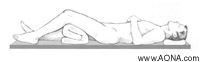
Position the patient
Position the patient supine on a radiolucent table. The knee of the injured leg should be flexed 30°-40° A leg roll may be used to allow for proper reduction and stabilization of the fracture. Ensure that x-ray visualization of the entire femur is possible in both the AP and lateral views
Click on image for enlarged view
Reduce the fracture. Use of the Large Distractor [394.35] is optional.
Intra-articular fractures should be stabilized with inter- fragmentary screw fixation prior to insertion of the nail. The screws should be positioned so as not to interfere with the path of the nail.
Position the ruler
Click on images for enlarged views
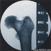
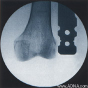
Position the image intensifier for an AP view of the distal femur. With a long forceps, hold the Radiographic Ruler [357.111] along the lateral aspect of the leg, parallel to and at the same level as the femur. Adjust the ruler until the distal end is at the desired nail insertion depth. Mark the skin at that site.
Move the image intensifier to the proximal femur, replace the distal end of the ruler at the skin mark, and take an AP image of the proximal femur. Verily fracture reduction. Read nail length directly from the ruler image, selecting the measurement that places the proximal end of the nail at the level of the lesser trochanter.
Note: All fractures should be treated with the longest nail possible, taking into account patient anatomy or previous implant.