|
|
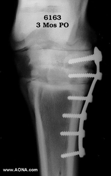
Radiographs taken 3 months postoperatively show all implants intact, and ongoing bony bridging of the distal intertarsal joint. (See Figure 4). The horse has been through a carefully prescribed exercise program including passive manipulation and controlled exercise, and is at this point being ridden at the walk, with occasional jogging to check degree of soundness. No lameness apparent.
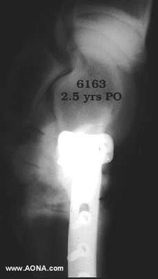
The horse continued on a problem-free course of recovery, and was performing well at intermediate level dressage two and one-half years postoperatively. The implants were never removed. (See Figure 5). The unfortunate proximal to distal angulation of this referral follow-up somewhat distorts the relationships of implant to bone, but 80-90 percent obliteration of the distal intertarsal joint space without excessive periosteal new bone is documented.
Conclusion: Plate arthrodesis of the distal intertarsal joint may be used to successfully treat chronic osteoarthritic lesions of this small tarsal articulation. Postoperative pain is minimal, and a controlled exercise program may be instituted 2 weeks postoperatively, and continue for the ensuing 10 weeks. Long-term results are effective, and aesthetically pleasing.